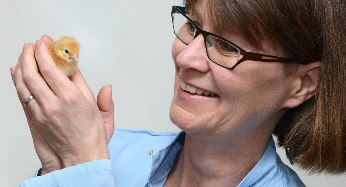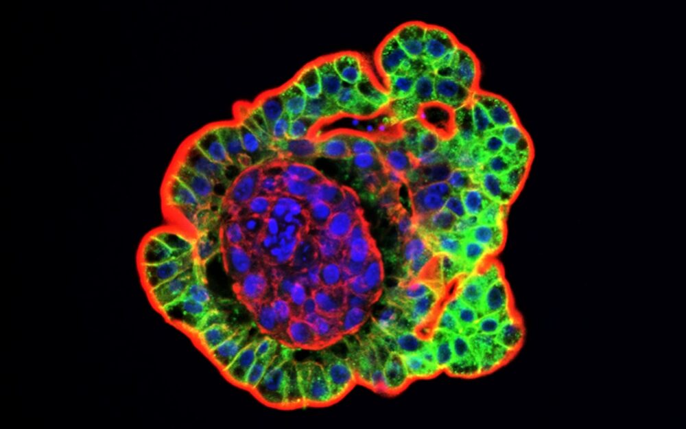
Professor Lonneke Vervelde’s inverted 3D chicken gut organoid model enables studies into poultry health while reducing the need for animal testing.
Complex in vitro cell culture systems that can better model in vivo biology is a rapidly developing field, driven by high drug attrition rate in drug development and the ethical concern surrounding the use of animal models. Particular market enthusiasm can be seen for organ-on-a-chip technology, but the whole market is expanding with associated downward price pressures on the technology.
Rising research and development in 3D cell culture technology, prominent presence of manufacturers, and initiatives for replacing animal models in research studies with organoids are some of the factors propelling the growth of the organoid market.
In vitro avian gastrointestinal studies have long been hampered by the lack of representative intestinal cell culture models. Existing 3D mammalian organoids lack components of the immune system and often present difficult access to the apical epithelium. Avian organoid models have so far yielded limited results.

Researchers at the University of Edinburgh’s Roslin Institute, led by Professor Lonneke Vervelde, Personal Chair of Veterinary Immunology & Infectious Diseases, developed a unique in vitro 3D inverted intestinal organoid model that mimics the chicken gut. They developed a complex avian organoid system that consists of multiple differentiated cell types present in the gut epithelium and in contrast to mammalian gut organoid systems, it also contains the immune and support cells from the underlying lamina propria. It overcomes drawbacks of previous models and differs from existing 3D mammalian organoids as it can be cultured freely in suspension without the aid of a structured support. One can manipulate the media in which the cells are floating by introducing pathogens, probiotics, vaccines etc, allowing researchers to use imaging and other recording to easily gauge how the gut reacts. The organoid has multiple villus-crypt structures that maintain the cellular diversity and barrier function present within the chicken intestinal epithelium, and its underlying lamina propria, in vivo.
This organoid, grown in a petri dish, is used for researching gut biology and the immune response to disease in chickens, as well as for testing environmental factors, feed additives, vaccines and drugs targeted at chicken health. This innovative organoid technology, featuring the internal gut surface on their exterior of the organoid, enables researchers to easily expose the tissue to disease-causing organisms and feed additives, and then run experiments and monitor the effects in a lab environment, without the need for complicated manipulation or microinjection.
The 3D in vitro organoid tissue cultures are composed of the chicken gut cell types and include cells from the immune system, enabling a more comprehensive understanding of the chicken’s response to infection.
This technology was developed by Professor Vervelde and her team as part of a collaborative project with an industry partner with funding from BBSRC and has been supported by Edinburgh Innovations.
Following a decade of research, this technology is poised to accelerate studies into gut health and diseases that affect birds around the world, while reducing the number of animals used in research. It also has an advantage for diagnostics of field samples that do not grow in cell lines, i.e. to isolate/discover new virus strains from the field.
Edinburgh innovations are now working with Professor Vervelde to support the further development of applications of this exciting new technology in a wide range of fields and with a diverse range of commercial partners. The project has also paved the way for a patent filing which is currently pending and will support the future commercial exploitation of the technology.
The applications will be particularly attractive to companies working in sectors such as nutrition, food additives, therapeutics, vaccines and infection control.
